The precise creation of nanoscale light fields is critical in emerging nanophotonics and nanooptics. Approaches for directly converting light into electrical impulses, along with non-destructive and accurate imaging of nanoscale light, may prove very beneficial.

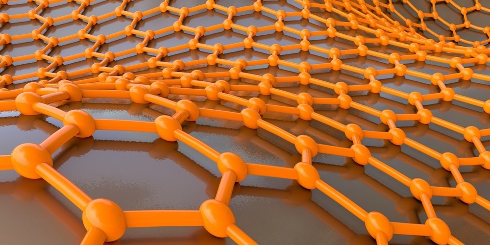
Study: Nanoscale light field imaging with graphene. Image Credit: Kateryna Kon/Shutterstock.com
In a study published in the journal Communications Materials a nanometric light field imaging technique was developed, centered around photodetection using a p-n junction which is generated and shifted by gate voltage within a graphene probe, generated by a series of exterior electrodes.
Current State of Nanophotonics
Nanoscale light modulation and characterization are two foundations of contemporary nanophotonics. Considerable developments in this subject have lately been made via the utilization of plasmonic components and their combinations, plasmon waveguides, super-lenses, optical phononics, and graphene-based plasmonics.
Meanwhile, for the most part, light field imaging and characterization still depend on microscopy approaches: measurement of dispersed propagation optical fields which are constrained to the diffraction threshold, restricting spatial resolution to a small portion of the complete light wavelength.
Near-Field Microscopy Methods for Improved Light Field Imaging
Using optical near fields may dramatically increase the accuracy of light field imaging. However, the basic six-power modulation of dispersed power using probe sizes in optical near-field microscopy restricts the practical resolution for direct light tracing to 50 nanometers at visible wavelengths.
Since the mapped optical fields are dispersed by a probe submerged in an examined optical near field, conventional near-field microscopy is thus essentially invasive.
Scattering scanning near-field optical microscopy (s-SNOM) having interferometric pseudo-heterodyne detection, the powerhouse of contemporary near-field optical characterization, uses demodulation of received signals at elevated harmonics to subdue the background noise. This is a rather effective strategy; however, it generates distortions in the imaging when fields with various spatial frequency bands are examined.
Optical Vs. Electron Microscopies
Different electron microscopy methods, like cathodoluminescence (CL) microscopy and electron energy-loss spectroscopy (EELS), have been practiced for obtaining details on the optical behavior of nanoscale structures with unrivaled, sub-nanometer level, spatial precision by employing highly focused beams of electrons.
Examining spatial mappings of matching EELS or discharged radiation bands, measuring the efficacy of resonance excitations of hybrid polariton forms, and linking the efficacy measurements with spatial distributions of the polariton forms yields the optical details.
Thus, electron microscopy approaches provide an indirect route to optical details, which must be extracted using complicated data processing techniques, primarily those connected with resonance excitations.
Indirect Light Imaging Methods
Indirect light imaging technologies based on dispersed fields may potentially attain extraordinarily fine spatial precision up to one nm.
For instance, tracing of a pigment particle's Brownian motion was performed to capture the fluorescence signature of a singular hot spot with one nm resolution. Surface-enhanced Raman spectroscopy (SERS) may be employed to assess electric fields with a five nm spatial resolution, whilst backscattered radiation may be utilized to detect optical oscillatory trends of plasmonic modes with a ten nm resolution using s-SNOM.
Unfortunately, despite offering incredibly high spatial resolutions, such indirect approaches are often invasive and depend on specific presumptions that enable one to convert observed values to light field mappings.
Highlights of the Study
In this study, the team proposed and tested electromagnetic field projections predicated on nanophotonic detection using a p-n junction produced and shifted within graphene by an externally applied gate voltage.
The p-n junction breadth of 20 nanometers determined the spatial precision of the electrical scanning approach, which may be increased even more by lowering the gate dielectric thickness.
An exterior gate voltage may precisely regulate the location of this kind of a p-n junction. The optical field mapping plane is defined by the graphene surface, which should be positioned near a nano-sized structure producing nanoscale light fields for characterization.
The proposed method was illustrated by tracing the electric field spread of a heavily constrained plasmon slot-waveguide mode at telecommunication wavelengths, yielding a mode profile that matched numerical predictions perfectly.
Notably, the unique arrangement demonstrated strong electro-optical modulating properties, with a 0.12 dB μm-1 modulating depth at a 6 V gate voltage amplitude.
The non-invasive light mapping technique introduces a new perspective in nanophotonic characterization, ensuring extraordinarily high spatial resolution and accuracy while also presenting exciting potential for nanoplasmonic on-chip systems.
References
Yu, T., Rodriguez, F. et al. (2022). Nanoscale light field imaging with graphene. Communications Materials. Available at: https://doi.org/10.1038/s43246-022-00264-0


 Add Category

